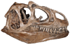
opis kręgów szyjnych z Calvo i in., 2007 B (zob. też ryciny) pisze:The atlas is one of the best preserved of any known
Titanosauria (Fig.3). The articulation with the
occipital condyle is wider than high. In lateral view,
the neural arch is displaced posteriorly (Fig.4). The
neurapophyses is a thin quadrangular lamina that
expands upward and curves medially, with the
distal end directed posteriorly. There is no contact
between both neurapophyses at the midline.
The axis has a short and high neural arch (Figs.5-
6). It occupies 2/3 of the total height of this element.
The odontoid process has not been preserved. The
neural spine is high, robust, of triangular shape. The
centrum is elongated without pleurocoels, differing
from Saltasaurus (POWELL, 1986) and Alamosaurus
(LEHMAN & COULSON, 2002). Prezygapophyses were not
preserved and postzygapophyses have a horizontal
articulation.
All cervical vertebrae are opisthocoelous with the
neural spines not bifurcated. Anterior cervical
elements are longer than high (Fig.7). The
triangular neural spine is robust and directed
posteriorly. The third cervical vertebra has robust
spinoprezygapophyseal and spinopostzygapophyseal
laminae and a smooth channel is developed
between them (Fig.8). On the fourth cervical, a deep
channel between both spinoprezygapophyseal
laminae is present, a feature observed in the
following elements of the neck. This channel does
not reach the top of the neural spine as observed
in titanosaurid cervical sequence from Brazil
known in the literature as the series A (POWELL,
1987), that latter received the number MCT 1487-
R (CAMPOS & KELLNER, 1999). The neural spine has
a triangular shape, in lateral view, and it is compressed
lateromedially but elongated anteroposteriorly
as the rest of anterior cervical vertebrae.
Pleurocoels are absent in all elements of the series,
a feature observed in Malawisaurus dixeyi and in
the sole cervical element known from Gondwanatitan
faustoi, respectively from Malawi and Brazil (JACOBS
et al., 1993; KELLNER & AZEVEDO, 1999). Parapophyses
are laminar and restricted to the anterior portion of
the centrum. The posterior centrodiapophyseal
lamina is directed anterodorsally as in MCT 1487-
R (POWELL, 1987) and it is different to that present in
Saltasaurus loricatus (BONAPARTE & POWELL, 1980).
Anterior cervical vertebrae of Titanosauria are scarce
in the fossil record, limiting further comparisons.
Middle cervical vertebrae are higher than long
(Fig.9). The centrum lacks pleurocoels as in MCT
1487-R from Brazil, but differing from the
condition reported in Malawisaurus and the
shallow lateral pleurocoels reported by CURRY
ROGERS & FORSTER (2001) in Rapetosaurus krausei.
The prezygapophysis in Futalognkosaurus
reaches the anterior border of the centrum,
different from the condition present in MCT 1487-
R and in the Saltasaurinae. The neural spine is
very high and sail-shaped as in Malawisaurus and
Rapetosaurus. Futalognkosaurs shares with
Rapetosaurus higher neural arches in anterior
and middle cervical vertebrae, three times higher
than the centra. They extend over the complete
length of the centra and are directed backwards.
In lateral view, the spinoprezygapophyseal border
is straight and the spinopostzygapophyseal
margin is concave, a feature not observed in
other members of the Titanosauria (Fig.9). The
only taxa with similar sail-shaped neural
spine is Rapetosaurus but it has the
spinopostzygapophyseal border straight
proximally and slightly concave distally.
Moreover, in Rapetosaurus postzygapophyses are
placed at middle height of the neural arch, as
those present in Rinconsaurus caudamirus (CALVO
& GONZÁLEZ RIGA, 2003). In anterior view, the
spinoprezygapophyseal laminae are fused on the
distal end forming a deep suboval depression.
This feature resembles, in some way, that present
in middle cervicals of the titanosaurid MCT 1487-
R from Brazil (POWELL, 1987). However, in the
latter, neural spines are very low with a rugose
and wide distal end. Middle cervical vertebrae
have a deep depression formed between the
base of the neural spine and the
diapopostzygapophyseal lamina (Fig.10). In
ventral view, a deep depression is present on the
proximal end of the centrum between the
parapophyses. This depression is considered an
autopomorphy of Futalognkosaurus dukei.
Posterior cervicals are opisthocoelous with very
elongated centra (Fig.11). Neural arches are high,
being three or more times higher than the centrum,
character only shared with Mendozasaurus neguyelap
(GONZÁLEZ RIGA, 2003). Neural spines are compressed
proximodistally and expanded laterally as in
Puertasaurus reuili (NOVAS et al., 2005) and in
Mendozasaurus, but to a lesser degree (Figs.11-12).
This shape is completely different in all other
titanosaurids such as Saltasaurus, MCT 1487-R from
Brazil, and Isisaurus colberti (JAIN & BANDYOPADHYAY,
1997). The neural spine is inclined slightly posteriorly,
different from the condition reported in Isisaurus
colberti, Puertasaurus reuili, and Mendozasaurus
neguyelap that are perpendicular to the body axis. It
displays an intraprezygapophyseal lamina and deep
supradiapophyseal cavities as those present in
Isisaurus and Mendozasaurus. In anterior view, no
prespinal lamina is present (Fig.12). In Isisaurus, a
true prespinal lamina is developed while in
Mendozasaurus the prespinal lamina is restricted to
the base of the neural arch (GONZÁLEZ RIGA, 2005).
Both spinoprezygapophyseal laminae in
Futalognkosaurus are robust and reach almost the
top of the neural spine (Fig.12). They are placed
almost parallel to each other, leaving a slit-shaped
depression between them. In Mendozasaurus and
Puertasaurus, the spinoprezygapophyseal laminae are
well separated and only reach the middle part of the
neural spine. Other Titanosauridae such as
Saltasaurinae (POWELL, 1986) and Rinconsaurini
(CALVO et al., this volume), also show this feature, but
the cavity is shallow. The last cervical vertebra (a
cervicodorsal), shows a prespinal-like lamina but it
does not reach the base of the neural arch. The
supradiapophyseal cavity is separated by a septum
from a lower depression placed on the diapophysis
(Fig.13). Futalognkosaurus dukei differs from the giant
titanosauriform Sauroposeidon proteles (WEDEL et al.,
2000) which has extremely elongated cervical centra
with a low neural arch, deep pleurocoels, and a deeply
excavated neural spine.


